Friendly Fire After the Infusion
Clinical Vignette
A 62-year-old man with recurrent HPV-positive oropharyngeal squamous cell carcinoma presents to the emergency department with two weeks of progressive exertional dyspnea and a dry, nonproductive cough. His cancer was treated definitively with cisplatin and radiation two and a half years ago, but pulmonary metastases appeared six months ago and he began pembrolizumab monotherapy; he has now completed eight cycles with a partial response. His other history includes a 40 pack-year smoking history with moderate chronic obstructive pulmonary disease, hypertension, and dyslipidemia.
Over the past two weeks his exercise tolerance has fallen from walking a block to becoming breathless while dressing, and for the last four days he has had low-grade fevers to 38.1 C, fatigue, and a 4-kilogram weight loss. He denies purulent sputum, hemoptysis, pleuritic chest pain, orthopnea, leg swelling, sick contacts, or recent travel. Two days ago his oncologist started empiric ceftriaxone and azithromycin for suspected community-acquired pneumonia, but his dyspnea has worsened and he now requires supplemental oxygen.
On examination, temperature is 38.0 C, heart rate 98/min, blood pressure 132/72 mmHg, respiratory rate 24/min, and oxygen saturation 89% on room air, improving to 95% on 6 L/min nasal cannula. He is chronically ill-appearing but not in shock. Lung examination reveals diffuse fine bibasilar inspiratory crackles without wheezes, dullness, or signs of consolidation. There is no lower extremity edema, no jugular venous distension, no calf tenderness, and no oropharyngeal pooling or dental infection. Laboratory studies show white blood cell count 11.2 x 10^3/uL with a normal differential and no eosinophilia, absolute eosinophil count 0.1 x 10^3/uL, hemoglobin 12.1 g/dL, creatinine 0.9 mg/dL, D-dimer 1.1 ug/mL, C-reactive protein 112 mg/L, and procalcitonin 0.06 ng/mL. Respiratory viral polymerase chain reaction testing, including SARS-CoV-2, influenza, and RSV, is negative, blood cultures are drawn, and serum beta-D-glucan and galactomannan are not detected. An echocardiogram shows normal left ventricular function.
CT pulmonary angiography demonstrates no pulmonary embolism but reveals bilateral, lower-lobe-predominant ground-glass opacities with fine reticulation and peripheral perilobular accentuation, without consolidation, cavitation, pleural effusion, nodules, or mediastinal lymphadenopathy. Because he is worsening despite antibacterial therapy, bronchoscopy is performed; bronchoalveolar lavage is negative for bacteria, mycobacteria, fungi, and respiratory viruses, cytology is benign, and the lavage differential shows a lymphocytic predominance. The imaging is shown below.
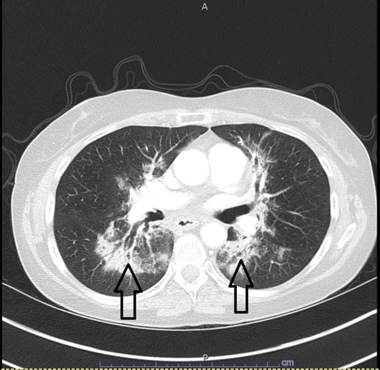
Axial CT of the chest: bilateral ground-glass opacities with fine reticulation — the classic radiographic pattern of checkpoint inhibitor pneumonitis.
Question 1
In this patient on pembrolizumab, what is the most likely diagnosis?
Select one option to submit your answer and view live poll results.
Question 2
Which statement best describes the diagnostic approach to this condition?
Select one option to submit your answer and view live poll results.
Question 3
After bronchoalveolar lavage returns negative, what is the most appropriate management of this patient's Grade 3 pneumonitis?
Select one option to submit your answer and view live poll results.
References
Naidoo J, Wang X, Woo KM, et al. Pneumonitis in Patients Treated With Anti-Programmed Death-1/Programmed Death Ligand 1 Therapy. Journal of Clinical Oncology. 2017;35(7):709-717.
Schneider BJ, Naidoo J, Santomasso BD, et al. Management of Immune-Related Adverse Events in Patients Treated With Immune Checkpoint Inhibitor Therapy: ASCO Guideline Update. Journal of Clinical Oncology. 2021;39(36):4073-4126.
Brahmer JR, Lacchetti C, Schneider BJ, et al. Management of Immune-Related Adverse Events in Patients Treated With Immune Checkpoint Inhibitor Therapy: ASCO Guideline. Journal of Clinical Oncology. 2018;36(17):1714-1768.
Haanen JBAG, Carbonnel F, Robert C, et al. Management of toxicities from immunotherapy: ESMO Clinical Practice Guidelines for diagnosis, treatment and follow-up. Annals of Oncology. 2017;28(suppl 4):iv119-iv142.
Habib T, Abu-Abaa M, Kolman-Taddeo D. Nivolumab-Induced Organizing Pneumonia in Management of Non-small Cell Lung Carcinoma: A Case Report. Cureus. 2023;15(5):e39217. [CT figure source — CC BY 4.0.]