Cut Once, Worse Twice
Clinical Vignette
A 34-year-old woman with ileocolonic Crohn disease presents with a rapidly enlarging ulcer on the right lower leg. Her Crohn disease has been difficult to control over the past year, with worsening diarrhea, abdominal cramping, weight loss, and a draining perirectal fistula. Fecal calprotectin measured two weeks before admission was greater than 3000 mcg/g. She had previously received adalimumab but stopped it eight months ago after losing insurance coverage. She is currently taking intermittent prednisone tapers and mesalamine. She has no diabetes, peripheral vascular disease, injection drug use, animal bites, freshwater exposure, or recent travel.
Three weeks before admission, she noticed a tender pustule on the anterior right shin that she attributed to minor trauma from bumping the leg against a coffee table. Over several days it opened into a painful ulcer with surrounding erythema. She was treated in urgent care with oral cephalexin, then trimethoprim-sulfamethoxazole, without improvement. She presented to the emergency department when the lesion doubled in size over 48 hours and developed a dark necrotic-appearing center. Initial concern was for a complicated skin and soft tissue infection. Vancomycin and piperacillin-tazobactam were started, and surgical debridement was performed.
The wound worsened after the procedure. The debrided margins became more violaceous and undermined, and new satellite pustules appeared along the incision line. Over the next week she underwent two additional debridements for presumed uncontrolled infection, each followed by further enlargement of the ulcer. Blood cultures remained negative. Operative Gram stain showed many neutrophils but no organisms, and tissue cultures grew only rare coagulase-negative staphylococci from enrichment broth. Fungal and acid-fast bacilli stains were negative. CT angiography of the leg showed soft tissue edema but no gas, drainable abscess, arterial occlusion, or osteomyelitis.
On infectious diseases consultation, she is afebrile with heart rate 94/min, blood pressure 124/76 mmHg, and oxygen saturation 99% on room air. The right anterior shin has a 9-cm ulcer with a purulent-appearing base, violaceous undermined borders, exquisite tenderness, and surrounding erythema that is most intense at the wound edge. There is no crepitus, bullous hemorrhage, anesthesia, lymphangitic streaking, or pain extending beyond the visible border. Laboratory studies show white blood cell count 12.4 x 10^3/uL, hemoglobin 10.1 g/dL, platelet count 520 x 10^3/uL, C-reactive protein 118 mg/L, erythrocyte sedimentation rate 88 mm/h, creatinine 0.7 mg/dL, and albumin 2.8 g/dL. Dermatology performs an incisional biopsy from the active ulcer edge. Histopathology shows a dense dermal neutrophilic infiltrate with sterile abscess formation and no vasculitis, granulomas, organisms, or malignancy. The lesion is shown below.
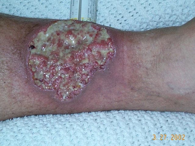
Lower leg ulcer with surrounding erythema and violaceous undermined borders in a patient with Crohn disease. Image: Crohnie, Wikimedia Commons, public domain.
Question 1
What diagnosis best explains this patient's progressive right lower extremity lesion?
Select one option to submit your answer and view live poll results.
Question 2
Which diagnostic approach is most appropriate once pyoderma gangrenosum is suspected?
Select one option to submit your answer and view live poll results.
Question 3
What is the best management strategy now that biopsy supports pyoderma gangrenosum and necrotizing infection has been reasonably excluded?
Select one option to submit your answer and view live poll results.
References
Maverakis E, Ma C, Shinkai K, et al. Diagnostic Criteria of Ulcerative Pyoderma Gangrenosum: A Delphi Consensus of International Experts. JAMA Dermatology. 2018;154(4):461-466.
Ormerod AD, Thomas KS, Craig FE, et al. Comparison of the two most commonly used treatments for pyoderma gangrenosum: results of the STOP GAP randomized controlled trial. BMJ. 2015;350:h2958.
Partridge ACR, Bai JW, Rosen CF, Walsh SR, Gulliver WP, Fleming P. Effectiveness of systemic treatments for pyoderma gangrenosum: a systematic review of observational studies and clinical trials. British Journal of Dermatology. 2018;179(2):290-295.
Peyrin-Biroulet L, Van Assche G, Gomez-Ulloa D, et al. Systematic Review of Tumor Necrosis Factor Antagonists in Extraintestinal Manifestations in Inflammatory Bowel Disease. Clinical Gastroenterology and Hepatology. 2017;15(1):25-36.e27.