Contaminant
Clinical Vignette
A 34-year-old man with HIV diagnosed two months ago presents to the infectious diseases clinic with three weeks of fever, productive cough, night sweats, and 6 kg of unintentional weight loss. His CD4 count at diagnosis was 42 cells/uL with a viral load of 280,000 copies/mL. He has not yet started antiretroviral therapy. He lives in a rural area of central Texas and works as a landscaper. He has no recent travel, no known tuberculosis exposure, and no animal contact beyond a household dog.
On examination his temperature is 38.9 C, heart rate 104/min, blood pressure 118/72 mmHg, respiratory rate 18/min, and oxygen saturation 95% on room air. He appears chronically ill with temporal wasting. Crackles are present in the right upper lung field. There is no lymphadenopathy, skin lesions, or organomegaly.
Laboratory studies show white blood cells 11.8 x 10^3/uL with 78% neutrophils, hemoglobin 10.2 g/dL, platelets 285 x 10^3/uL, creatinine 0.9 mg/dL, ALT 42 U/L, and AST 56 U/L. Three induced sputum specimens for acid-fast bacilli smear and mycobacterial culture are all negative. A fourth sputum shows no AFB. Histoplasma urine antigen is negative. Serum cryptococcal antigen is negative.
Chest CT demonstrates a 4.5 cm cavitary lesion in the right upper lobe with a thick irregular wall and surrounding parenchymal consolidation. There is no pleural effusion or mediastinal lymphadenopathy. The radiologist's impression notes that the findings are concerning for tuberculosis or fungal infection in the setting of advanced HIV.
Bronchoscopy with bronchoalveolar lavage is performed. Bacterial culture of the BAL fluid grows a gram-positive coccobacillus after 48 hours. The microbiology laboratory initially reports it as "Corynebacterium species, probable skin contaminant." However, the laboratory's modified Kinyoun (modified acid-fast) stain on the direct BAL specimen is positive, showing organisms that retain carbolfuchsin with 1% sulfuric acid decolorization. On blood agar, the colonies are salmon-pink, smooth, and mucoid. The organism is catalase-positive and urease-positive.
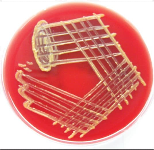
Blood agar culture after 48 hours of incubation demonstrating pigmented mucoid colonies. Image from Dias et al., J Family Med Prim Care, 2013, CC BY-NC-SA 3.0.
Question 1
What is the most likely causative organism?
Select one option to submit your answer and view live poll results.
Question 2
Which virulence mechanism best explains why this organism causes opportunistic infection in patients with advanced HIV?
Select one option to submit your answer and view live poll results.
Question 3
Which approach to treatment is most appropriate?
Select one option to submit your answer and view live poll results.
References
Prescott JF. Rhodococcus equi: an animal and human pathogen. Clinical Microbiology Reviews. 1991;4(1):20-34.
von Bargen K, Polidoro M, Klose K, et al. R. equi VapA is required for diversion of phagosome biogenesis but not for cytotoxicity. Infection and Immunity. 2009;77(12):5676-5681.
Harvey RL, Sunstrum JC. Rhodococcus equi infection in patients with and without human immunodeficiency virus infection. Reviews of Infectious Diseases. 1991;13(1):139-145.
Topino S, Galati V, Grilli E, Petrosillo N. Rhodococcus equi infection in HIV-infected individuals: case reports and review of the literature. AIDS Patient Care and STDs. 2010;24(4):211-222.
Weinstock DM, Brown AE. Rhodococcus equi: an emerging pathogen. Clinical Infectious Diseases. 2002;34(10):1379-1385.