The Daughter Within
Clinical Vignette
A 34-year-old woman from rural Patagonia, Argentina, who has lived in the United States for six years presents to her primary care physician with three months of intermittent right upper quadrant fullness and mild discomfort. She grew up on a sheep farm where her family kept working dogs that herded livestock and had free access to the animals' viscera at slaughter. She recalls no acute illness during childhood.
On examination she is afebrile, hemodynamically stable, and has mild right upper quadrant tenderness without guarding or palpable mass. Liver function tests show total bilirubin 0.7 mg/dL, alkaline phosphatase 98 U/L, ALT 42 U/L, and AST 38 U/L. Complete blood count and basic metabolic panel are unremarkable, with a normal eosinophil count.
Abdominal ultrasound reveals a 12 cm well-defined cystic lesion in the right hepatic lobe with multiple smaller peripheral cystic structures within it, creating a characteristic spoke-wheel or rosette pattern. No biliary ductal dilatation is seen. Contrast-enhanced CT of the abdomen confirms a 12.4 x 10.8 cm multiloculated cystic lesion in hepatic segments VII and VIII containing numerous rounded daughter cysts of varying size along the periphery, with a thin curvilinear calcification along portions of the wall. The cyst wall does not enhance after contrast administration. No solid enhancing component is identified. The remainder of the liver, biliary tree, and pancreas are normal.
Echinococcus IgG ELISA returns positive. Western blot confirms specific reactivity with the arc 5 antigen. The surgical team plans CT-guided percutaneous aspiration of the cyst for definitive histopathologic diagnosis.
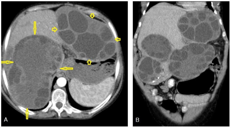
Contrast-enhanced CT abdomen demonstrating a large multiloculated hepatic cyst with characteristic peripheral daughter cysts in a spoke-wheel pattern. Image from Alshoabi et al., Diagnostics, 2023, CC BY 4.0.
Question 1
What is the most likely diagnosis?
Select one option to submit your answer and view live poll results.
Question 2
The surgical team proposes CT-guided percutaneous aspiration of the cyst for histopathologic diagnosis. What is the most important concern with proceeding?
Select one option to submit your answer and view live poll results.
Question 3
According to the WHO-IWGE stage-specific management framework, which treatment approach is most appropriate for this patient's cyst?
Select one option to submit your answer and view live poll results.
References
Brunetti E, Kern P, Vuitton DA. Expert consensus for diagnosis and treatment of cystic and alveolar echinococcosis in humans. Acta Tropica. 2010;114(1):1-16.
World Health Organization. WHO guidelines for the treatment of patients with cystic echinococcosis. Geneva: WHO; 2025.
Alshoabi SA, Al-Zahrani HM, Baessa EM, et al. Hydatid disease: a radiological pictorial review. Diagnostics. 2023;13(6):1127.
Stojkovic M, Zwahlen M, Teggi A, et al. Treatment response of cystic echinococcosis to benzimidazoles: a systematic review. PLoS Neglected Tropical Diseases. 2009;3(9):e524.
Neumayr A, Troia G, de Bernardo C, et al. Justified concern or exaggerated fear: the risk of anaphylaxis in percutaneous treatment of cystic echinococcosis. PLoS Neglected Tropical Diseases. 2011;5(6):e1154.