The Copper Penny Sign
Clinical Vignette
A 47-year-old male farmer from the state of Pará in northern Brazil presents with a three-year history of a slowly enlarging skin lesion on his right lower leg. The lesion began as a small, firm papule after a minor scratch from a wooden fence post and has progressively enlarged over the years. He reports no pain, fever, or systemic symptoms. He has not sought medical attention previously, attributing the lesion to a chronic wound from his agricultural work.
On examination the lesion occupies a 12 by 9 cm area of the right lower leg. It is a raised, verrucous, cauliflower-like plaque with a hyperkeratotic surface, irregular borders, and scattered crusted areas. The surface has a dark gray-brown discoloration. There is no fluctuance, warmth, or significant surrounding erythema. Regional lymph nodes are not enlarged. The remainder of the examination is unremarkable; there are no pulmonary, hepatic, or neurologic findings.
A punch biopsy of the lesion is performed. The tissue is sent for histopathology and fungal culture on Sabouraud dextrose agar. The histopathologic result is shown below.
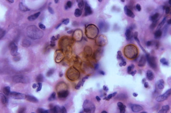
Skin biopsy, hematoxylin and eosin stain: brown, thick-walled, spherical to polyhedral muriform cells. Image courtesy of the CDC.
Question 1
The biopsy shows brown, thick-walled muriform cells with transverse septa clustered within the dermis. What is this pathognomonic structure called, and what diagnosis does it confirm?
Select one option to submit your answer and view live poll results.
Fungal culture of the biopsy on Sabouraud dextrose agar yields a slowly growing colony over three weeks. The colony is dark olive-green to black, with a velvety surface and a black reverse. Microscopic examination of the culture is shown below.
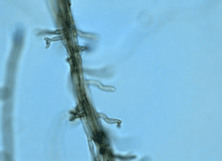
Lactophenol cotton blue preparation: dark brown, septate, loosely branching hyphae with pale brown to olivaceous conidia. Multiple conidiation types are present, including Rhinocladiella-type, Cladosporium-type, and Phialophora-type patterns.
Question 2
Culture yields a slow-growing, dark olive-black colony with the microscopic morphology shown. Which organism is the most likely cause of this patient's chromoblastomycosis?
Select one option to submit your answer and view live poll results.
Question 3
The diagnosis of chromoblastomycosis is confirmed. What is the treatment of choice?
Select one option to submit your answer and view live poll results.
References
Queiroz-Telles F, de Hoog S, Santos DW, et al. Chromoblastomycosis. Clinical Microbiology Reviews. 2017;30(1):233-276.
Queiroz-Telles F, McGinnis MR, Salkin I, Graybill JR. Subcutaneous mycoses. Infectious Disease Clinics of North America. 2003;17(1):59-85.
Seyedmousavi S, et al. Scope and challenges of emerging and uncommon fungal diseases in Europe: leading clinicians from across Europe provide their view. Lancet Infectious Diseases. 2018;18(11):e344-e352.